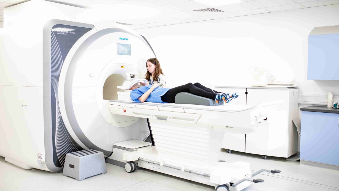
Imaging and biosensing technologies for objective decision-making
Clare Green, Technology Innovation Manager at Grow MedTech, explores monitoring and diagnostic tools being developed in our partner universities.
As the new era of personalised medicine gets underway, doctors are requiring increasingly sophisticated and intelligent decision-making and monitoring tools. These diagnostic tools will help them make treatment decisions that are relevant to highly specific subsets of patients, or even personalised to an individual.
From wearable monitoring devices, to smart diagnostics that use artificial intelligence (AI) and machine learning (ML) to learn and adapt, these new technologies are becoming essential to help doctors spot conditions early and deliver the right treatment, at the right time.
The potential benefits to patient wellbeing, as well as the savings in NHS resources, are significant.
At Grow MedTech, we support researchers to evidence how their technology developments will provide such savings.
Our Proof of Market and Proof of Feasibility funds can help researchers move projects through the early development stages, where it is often hard to find funding, but where being able to demonstrate aspects such as market need and health economics is crucial for moving to the next Technology Readiness Level.
Solving the microwave challenge
The University of York has particularly impressive capabilities in the field of imaging, biosensing and diagnostics technology, with cutting-edge research being carried out in a number of departments and access to facilities such as its Centre for Hyperpolarisation in Magnetic Resonance (CHyM) and the Bioscience Technology Facility.
These facilities put the University in a great position to take advantage of our support.
One area in which York is excelling is in the use of microwave technology in diagnostics – specifically to measure the depth and severity of burns.
A team led by Professor Roddy Vann, in the York Plasma Institute, is currently working on a prototype device that can provide a 3D image of temperatures up to 2cm below the skin by measuring microwaves naturally emitted by the body, the strength of which depends on the temperature of the tissue.
This is expected to greatly advance research into burn progression through direct imaging of the damaged area.
Microwave imaging of burns has been considered in scientific literature since the 1970s but technical challenges have so far limited its translation into the clinic.
This device is innovative in combining recent advances in the design of antennas and ultra-fast data acquisition with the potential to make microwave medical imaging both technically and commercially viable.
We are helping to de-risk the project by demonstrating the feasibility of the prototype and supporting the team to develop the mathematics and software that will convert the complex signals that are emitted from the burn into a usable image that can be used by healthcare professionals to judge its severity
Scientists from industry partner, Sylatech, are co-inventors of the technology, working alongside Grow MedTech and the York team to commercialise the technology.
Working closely with industry from an early stage is always important and the link with Sylatech is particularly strong, having grown from its early days as a Knowledge Transfer Partnership.
We’ve also been fortunate to bring in clinicians from specialist burns centres at the Queen Elizabeth Hospital in Birmingham and Pinderfields Hospital in Wakefield – as well as Patient and Public Involvement groups – to guide this research.
Improving c-section infection diagnosis
Measuring the body’s heat signals is a promising area for diagnostics, and one in which Sheffield Hallam researchers are also focusing their attention with our support.
This time, infra-red thermal imaging cameras are being employed to help with decision-making, by predicting the likelihood of surgical site infection in women after Caesarean section.
Around 200,000 women undergo caesarean sections each year. Since it is hard to tell, by looking at a wound, whether or not it will become infected, women are routinely prescribed prophylactic antibiotics.
Identifying infections early and treating them appropriately will reduce patient risk, and cut down the number of antibiotics prescribed.
In the University’s Centre for Health and Social Care Research, Professor Charmaine Childs’ team is using Grow MedTech funds to investigate the potential market for the device before making a bid for significant national funding to refine the design and software ahead of a clinical trial.
In this way, our support is helping to bridge the gap between early stage research and more advanced technology development.
Objective disease diagnosis
One area in which diagnosis is particularly difficult is in diseases marked by cognitive decline, such as Alzheimer’s.
There are a number of challenges, including finding reliable biochemistry tests that can accurately measure changes, and obtaining the right samples from a particularly vulnerable group of patients.
At Leeds Beckett, we funded a Proof of Market project, led by Dr Nat Milton, to progress a non-invasive test based on saliva samples. Working with the University of Huddersfield and the University of Roehampton, Dr Milton’s team has identified biochemical markers called kisspeptins that are found in saliva and can be linked to Alzheimer’s.
We worked with Dr Milton to support a detailed exploration of the potential market for this test, along with an early stage cost benefit analysis. The results were extremely positive, showing a clear market opportunity for a test of this type.
The next step for the team will be able to finalise its development and produce a prototype device. It’s clear that there’s an urgent need to be able to diagnose Alzheimer’s more accurately and at an earlier stage and so it’s particularly exciting to see this technology progress towards the clinic.

