Interventional X-ray expert seeks new test tool
Translate MedTech’s Secondment Scheme is designed to develop the innovation skills and translational capability of medical technology researchers in the Leeds City Region. This blog is part of a series that showcases the impact that secondments have had on medtech research, as recounted from the secondee’s perspective.
Due to the success of the scheme, it is being run again in 2020. To find out how to apply for Translate secondment funding for your medtech research, click here.
Name: Dr Amber Gislason-Lee
Host organisation during the secondment: Guys and St Thomas' Trust
(London) and UZ Leuven (Belgium)
My name is Dr Amber Gislason-Lee and I teach medical imaging science and technology to radiography students at the University of Bradford’s School of Allied Health Professionals.
My background is in medical physics, which puts me in a unique position to teach this topic and to contribute to research in medical imaging.
I previously worked under the mentorship of Arnold Cowen, who designed the original Leeds Test Objects before they were bought by a commercial company outside of Leeds.
One area of research which I have many years of experience in is interventional X-ray imaging. For that reason, I am on a working group for the Institute of Physicists and Engineers in Medicine (IPEM) to write a quality assurance (QA) guidance document for medical physicists testing interventional X-ray systems.
This guidance document has been challenging for the group to write, one of the main reasons being a lack of X-ray test object to mimic the motion of a human heart, to check for temporal and spatial resolution of the X-ray system at hand.
These aspects of image quality allow for better accuracy in treating patients via angioplasty, radiofrequency ablation and other life-saving interventional cardiovascular procedures. Since joining the University of Bradford last year I have discovered the collaborative potential between radiography and engineering and reached out to some engineering colleagues to discuss potential projects to address ongoing X-ray research problems to be solved.
One of the results of these was a collaborative relationship with Peter Twigg from Engineering. After a successful application for medtech funding to support a summer student engineering project, we had a prototype X-ray test object which addressed the needs brought to light by the IPEM group.
Although I found the prototype seemed potentially useful and affordable for hospital physics departments, I needed to run it by my colleagues who have more day-to-day testing experience than me. I am an academic involved with teaching and research in medical physics and they are hospital physicists doing routine testing and troubleshooting, so there is a big difference between these roles.
Given their busy schedules we made arrangements for a short meeting at the end of the workday (in hopes the patients would be finished with the X-ray machines) – only a few hours long. This secondment allowed for the funding of travel to these meetings.
Test Object well-received at visit 1 of 2
There are two key medical physics experts whom I sought to visit for this secondment, to bring the test object prototype to them in person, show them how it works and potentially view it under X-rays should an interventional X-ray system be available for use.
Ian Honey works as a hospital physicist at Guy’s and St Thomas’ Trust in London, UK. Nick Marshall, formerly of St Barts Hospital, London, works at UZ Leuven in Belgium and is now an active member of the European medical physics community.
Both these experts have written IPEM QA guide books previously, and both have expressed their desire for a test object such as the one designed at Bradford. I should clarify that during my previous conversations with these experts they communicated what was required of the test object, however, they did not have any specific ideas for its design and how it would achieve its goals.
The design by Peter Twigg is very unusual, different than any of the Leeds Test Object designs that physicists around the world are accustomed to using. For this reason, I did not provide any details about the test object prior to arriving on-site and opening the box. I only divulged its uniqueness.
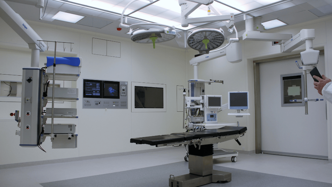
In London, Ian had 2 colleagues with him as well as a (dated) X-ray system to use for testing. The comments were very positive from all 3 physicists, generally foreseeing the test object as a troubleshooting tool for situations where clinical X-ray systems have had temporal blur issues reported. We spent a few hours testing the object using a range of clinically relevant experimental setups, and I captured both stills and videos on my phone (see attached).
We all enjoyed ourselves and it made me proud to bring our design form the North to such a prestigious London hospital. Ian and Nick are very different from each other, though both very honest – so although I was very excited about the positive response from Ian I was very keen to see what Nick had to say before I let my excitement take off!
Moving forward with the product design
After my first meeting, I learned that it’s always worth ‘having a go’ at making something if you have the right people to do it! I was nervous about showing the test object to Ian, but it was well-reviewed. This is something I had never done before and it required a certain level of confidence in my own level of expertise in my subject area.
Nick, for very different reasons than Ian, also supported the test object design and could see its usefulness should some further testing be done on a modern interventional X-ray system. He offered to do these tests at his site in Belgium in the coming months.
Nick is one of a handful of medical physicists in the world who have access to unprocessed X-ray image data from an imaging system manufacturer, and this is a necessity for test object testing/validating. Even if Nick hadn’t liked it at all that would have been useful information to determine our next steps with the prototype – but what a fantastic result!
My next step is to meet with Peter and determine what our next steps should be to complete the design with the intent of turning the prototype into a commercially available product for medical physicists to purchase for use at their hospitals.
We also plan to show the test object to the entire interventional X-ray physics community at a national meeting on 1st May. The organiser of the meeting has put aside a time for Peter to bring the test object to the meeting in the hopes of receiving more detailed feedback from its potential users to feed into specific aspects of its final design.
There are 2 reasons why I really appreciate this secondment:
- I felt there was a definite need to show this test object to these 2 experts prior to anyone else and to do it IN PERSON. The travel was required in order to determine their initial reaction, and collect their suggestions for improvements and features to include in the design.
- I have no experience with ‘inventing’ a product, however, I feel I was well-positioned to communicate the requirements of the product. I have gained confidence in developing medical technology to address issues that I understand.
I would recommend this experience to anyone in a similar position – go for it!
_____________________________________________________________________________________________________________
If you could benefit from support while progressing a medical technology towards commercialisation, or if you want to develop innovation skills then consider applying for a Translate MedTech secondment.
Our latest call is open to applications, and further detail can be found on our website here. You can also contact Mohua Siddique with any questions you might have about the scheme.